represent EMBL in Europe.
Tissue biology and disease modelling
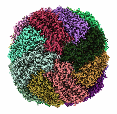
Structural biology
Main laboratory
EMBL-EBI: European Bioinformatics Institute
Epigenetics and neurobiology
Examples: 1001, Apoferritin, Tomography, Rossmann MG, 5A1A
advanced search
Current Database
Current status
Sample type
Organism
EM Method
Resolution
Model molecular weight
Release Date
Author by name
Author by ORCID
Journal
Software
Microscope
Electron Source
Film or detector model
External links
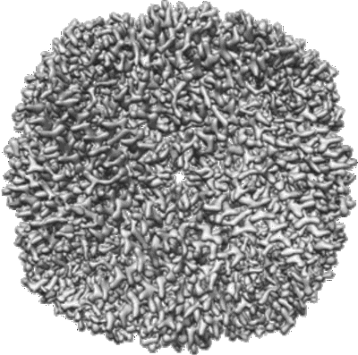
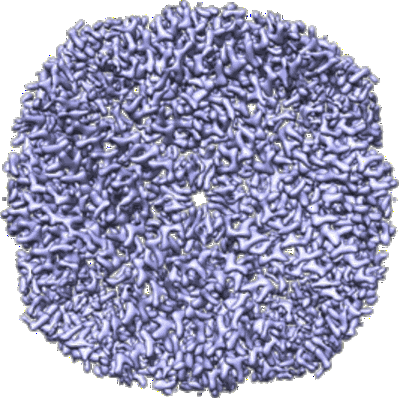
1.42 Angstrom Apoferritin structure determined using G1 Titan krios S-FEG operated at 300kV, zero loss imaging using Gatan BioQuantum energy filter operated at 10eV slit width and imaged using K2 camera.
Venugopal H, Ramm G
To Be Published
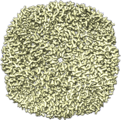
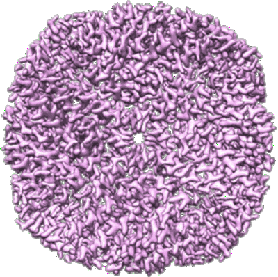
1.96 A structure of human apoferritin obtained from Talos Arctica microscope
Fan H, Wang B, Zhang Y, Zhu Y, Song B, Xu H, Zhai Y, Qiao M, Sun F
Nat Commun (2021) 12 pp. 7257-7257 [ DOI: doi:10.1038/s41467-021-27596-8 Pubmed: 34907237 ]
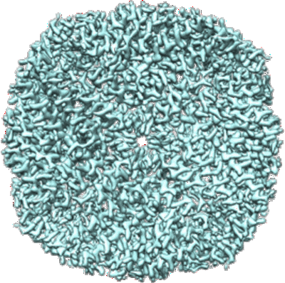
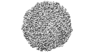
Apoferritin short exposure 3D reconstruction with 10% total dose
Bepler T, Kelley K, Noble AJ, Berger B
Nat Commun (2020) 11 pp. 5208-5208 [ Pubmed: 33060581 DOI: doi:10.1038/s41467-020-18952-1 ]
Apoferritin short exposure 3D reconstruction with 25% total dose
Apoferritin short exposure 3D reconstruction with 50% total dose
Apoferritin short exposure 3D reconstruction with 75% total dose
Apoferritin short exposure 3D reconstruction with 100% total dose
Apoferritin map obtained from grids prepared with the Preassis method
Zhao J, Xu H, Carroni M, Lebrette H, Wallden K, Moe A, Matsuoka R, Hogbom M, Zou X
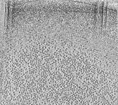
Apoferritin with spot-to-plunge time of 100ms
Noble AJ, Wei H, Dandey VP, Zhang Z, Tan YZ, Potter CS, Carragher B
Nat Methods (2018) 15 pp. 793-795 [ Pubmed: 30250056 DOI: doi:10.1038/s41592-018-0139-3 ]
Apoferritin with spot-to-plunge time of 400ms
Searching in EMDBHelp
Share EMDB